
-From Dr. Mohamed Omer Khider Ahmed
Gallbladder Polyps – are present in 4 to 6 percent of the population.4,5 An estimated 90% are benign cholesterol polyps, less than 10 mm in size and are incidental findings. The remaining 10% are adenomatous polyps that have malignant potential.
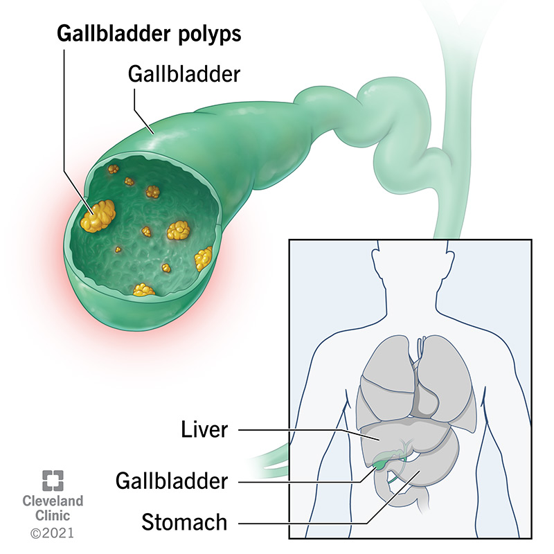
Most polyps are spherical (attached by a pedicle to the gallbladder wall). Less common are the broad based (sessile) polyps.
Sonographically a polyp appears as a hyperechoic nodule (more echogenic than the surrounding bile) attached to the gallbladder wall. The polyp is nonmobile and remains in a fixed position regardless of changes in patient position. The polyp is non-shadowing.
We have 36years old man sent to our clinic complain of right upper quadrant pain for an abdominal ultrasound, the study done using a handheld ultrasound devise from EagleView and gallbladder wall shows multiple immobile polypoid ingrowths into gallbladder lumen with no vascularity at color doppler which denotes gallbladder polyp.

image (1) sagittal view of the gallbladder demonstrated non-shadowing polypoid ingrowth into gallbladder lumen by the white arrow. gallbladder wall demonstrated by the black arrow.

image (2) transverse view of the gallbladder demonstrated non-shadowing polypoid ingrowth into gallbladder lumen by the white arrow. gallbladder lumen demonstrated by the black arrow.

image (3) color doppler over the gallbladder polyp (white arrow) demonstrated no color flow.
We are dedicated to developing products with medical-grade accuracy but consumer friendly, finding an easier and more modern way to track your wellness, we believe you can get peace of mind to live a healthier and longer life.