by welluehealth | Feb 23, 2022 | MED PROS
Since the American Heart Association’s recommendation to obtain prehospital 12-lead electrocardiograms on patients with acute coronary syndrome, EMS providers have played an increasingly important role in identifying these patients, beginning the appropriate treatment, and transporting them to appropriate hospitals capable of emergency angioplasty.
The acquisition of the 12-lead ECG in the field is theoretically not different from those obtained inside the emergency department. However, due to the unique prehospital environment, there are several tips and pearls to consider when placing the patient electrodes.
Conditions Requiring an ECG
A 12-lead isn’t just for chest pain. Obtain a 12-lead for possible strokes, altered levels of consciousness, weakness, dizziness, fatigue, palpitations and otherwise vague medical complaints. Remember that diabetic patient, younger women and various ethnicities often have atypical presentations and may have “silent myocardial infarctions.” Be vigilant. You may just save a life.
Proper acquisition of the 12-lead ECG
Proper acquisition of the 12-lead ECG is paramount to getting the most out of this tool. An improperly acquired 12-lead doesn’t provide diagnostic-quality information and can render the tracing mostly useless. Here are a few tips to making sure you get it done right:
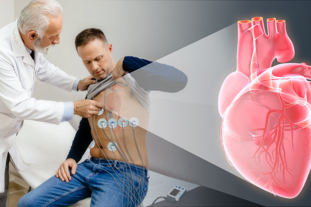
Skin Preparation
It’s important to prepare the skin by cleaning it with an alcohol prep and by abrading it with a cloth towel to remove dead skin cells. You may need to wash the area with saline and dry it. Remove excess body hair by shaving. For females, place the leads under the breast tissue. You may need to lift and clean the skin underneath the breast to get a clear tracing.
Lead Placement
Traditionally, the limb leads go on the limbs, and while it’s acceptable to move them closer if you have to, try to avoid placing the leads over bony prominences or overly fatty areas. Look for a generally flat, clean, intact area of skin with muscle generally underneath.
- The V-Leads go on the chest in a specific pattern. Leads V1 and V2 go in the 4th intercostal spaces (between the ribs) on either side of the sternum. To find these, go about three finger widths up from the xyphoid process, or bottom of the sternum. V1 is on the patient’s right, V2 is on the left.
- V4 should be placed next; it goes one rib down in the 5th intercostal space, on the midclavicular line. Place V3 in between V2 and V4.
- V5 goes in the anterior axillary line (front of the armpit) and V6 goes in the mid-axillary line. They go in the same horizontal line as V4.
Electrode Placement for Women
Asking a female patient to disrobe can be uncomfortable for the EMS provider, at first. Be professional. Explain to the patient what you plan to do in terms of electrode placement; emphasize that several of the chest leads may need to be placed around and under the left breast.
Have a patient gown available for the patient to use after removing her clothing.
If the patient’s left breast is large enough to cover the V3, V4, or V5 placement area, it will have to be lifted up for proper electrode placement. If possible, you can ask the patient to lift her own breast. Alternatively, use the back of your straightened hand to displace the breast.
Electrode Placement for Bariatric Patients
Obese patients may appear to be more difficult at first to accurately place electrodes. The trick is to spend a few extra moments locating the anatomic landmarks. Palpate more deeply to feel the sternal border and Angle of Louie to place leads V1 and V2. V4 is usually located in a straight line below the nipple at the fifth intercostal space. Then, imagine a line track straight down the left lateral side of the chest. Along this line, at the mid-axillary line is the location of lead V6.
Once these leads are placed, then V3 is placed halfway between V2 and V4. Finally, V5 is placed halfway between V4 and V6.
Electrode Placement for Pregnant Patients
Despite the appearance of the abdomen during advanced pregnancy, the placement of the electrodes is the same. You can use the technique above if necessary.
Note that left-axis deviation on the ECG may appear in both pregnant and obese patients. This is due to the abnormal position of the heart as the diaphragm pushes high into the thoracic cavity.
Electrode Placement for Pediatric Patients
Use smaller electrodes specific to children. Adult electrodes will overlap and potentially cause inaccurate placement. For preschool-age children and older, take time to explain what you are doing. Young children will be fearful of the procedure and may imagine that it will hurt, or that you will shock them. Having a parent close by will help provide reassurance.
Obtaining 12-Lead ECG in Extreme Environments
Extreme heat or cold will affect the integrity of the electrode’s conducting gel. During the cold winter or hot summer months, check to make sure that the electrode bag is kept in a location that minimizes dramatic temperature shifts.
Acquisition Tips to Minimize Artifact
Movement of any sort has the potential to create excessive artifacts in the ECG. Consider these tips:
If your patient is shivering, cover the skin with a light sheet and consider using a small heat pack to provide a sense of comfort. Turn the thermostat in the ambulance up to keep the patient warm.
The patient should be in a semifowler’s position.
Ask the patient to simply breathe normally and keep their hands by their sides. This prevents them from gripping the handrails too tightly, which can cause minute muscle tremors that show up on the ECG as artifacts.
There should be some “slack” in the patient cables. If the cable is taut between the electrode and the monitor, adjust the cable to release the tension.
With practice and preparation, obtaining a clean 12-lead ECG every time will be easier to accomplish. Your confidence in acquiring an accurate tracing will decrease the time it takes to decide how to manage and transport the patient who is experiencing ACS, and increase the chance of survival and recovery.
Baseline
A quality 12-lead ECG has a smooth, flat baseline (called the isoelectric line). Baseline wander, or the vertical motion of the ECG line, can mask important findings in the ECG tracing and result in a non-diagnostic ECG.
The patient should remain motionless and lay as close to supine as possible for the acquisition of the tracing and the ambulance should be stopped and not move during the process. It sometimes takes a few minutes for the ECG tracing to normalize and you should wait for it to do so. The goal is to be able to see the entire cardiac waveform clearly and be able to measure accurate ST-segment levels. Skin prep is important to reduce artifacts. A tracing with artifact or baseline wander can mask serious ECG findings and may cause a patient to be misdiagnosed.
We are dedicated to developing products with medical-grade accuracy but consumer friendly, finding an easier and more modern way to track your wellness, we believe you can get peace of mind to live a healthier and longer life.

by welluehealth | Feb 18, 2022 | MED PROS
Emergency medical service(EMS) is an essential part of the health care system and is always expected to be available always and continuously in case of need.
The 12-lead ECG is one of the most fantastic advances in EMS treatment since the invention of the bandage. It has proven to be a landmark addition to the EMS toolkit as the critical diagnostic aid to provide care in the field, guide transport to the most appropriate hospital for acute heart conditions, and allow notification of the hospital so that emergency staff can be prepared.
The movement of this powerful diagnostic tool from the confines of the hospital to the streets has been nothing short of revolutionary. It has given EMS professionals a wealth of information on how to best care for our patients and has driven hospital care and the development of medical care practices by providing clear and critical data that physicians had rarely before seen.
What is a 12-Lead ECG Machine?
A 12-lead electrocardiogram (ECG) is a medical test that is recorded using leads, or nodes, attached to the body.
It can identify a variety of heart problems, rhythm abnormalities, and cardiac injuries. Its addition serves as the basis for updating EMS protocols to improve the care of patients suffering many types of acute cardiac events.
Most importantly, the 12-lead EKG is the only tool to identify a patient with an ST-elevation myocardial infarction (STEMI). It could reproduce QRS, ST, and T waveforms accurately, and provides evidence of P wave and QRS complex morphology that cannot be determined by single-Lead ECG.
Not all hospitals provide acute treatment interventions for this condition, so the crucial EMS role is to identify patients with that disease and then, if possible, transport the patient for immediate intervention. There are many outstanding cases of EMS personnel identifying the patient having an MI, communicating that condition to the emergency department, and helping move the patient to the cardiac lab.
Application of 12-lead ECG in the diagnosis of STEMI
For ST-elevation myocardial infarction, the mortality of patients is closely related to the time from chest pain to perfusion (Ischemic to balloon time, also called myocardial hypoxia time).
The demonstrated advantages of 12-Lead ECG in EMS
In 2006, the New England Journal of Medicine published that if the cardiac catheterization room can be activated before the patient arrives at the hospital, it will effectively reduce the time of myocardial hypoxia. However, the proportion of patients who can complete the pre-hospital ECG diagnosis is still not high.
In some cities, ambulances are equipped with 12-lead ECG equipment. When the fire rescue arrives at the rescue scene, they can first perform an ECG examination according to the condition. If it is judged to be STEMI, then the catheter would be called to activate immediately. In this way, when the patient arrives at the hospital, the Cath Lab would be ready to arrange cardiac catheterization, so as to reduce the preparation time of the cath lab, thereby shortening the time from hospital to perfusion.
Actually, even if the ambulance directly transfers the patient to the cardiac catheterization hospital, the activation of the cath lab will take a lot of time, which includes the organization of the cardiac catheterization team, the preparation of machines and equipment, etc. At present, if the hospital needs to activate the acute cardiac catheterization team, the notification can only be confirmed after the patient arrives at the emergency room and complete the ECG diagnosis.
In the emergency scene, 12-Lead ECG is used to complete the ECG detection, and some of the machines are able to automatically interpret and transmit the ECG report to the expert to establish the diagnosis result in advance, and then transfer it to the cardiac catheterization hospital, thus the early treatment could reduce the time of myocardial hypoxia and mortality rate effectively.
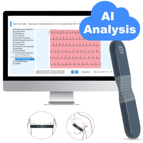
How Wellue can help
Wellue offers two types of portable 12-Lead ECG Machines, the Biocare 12-Lead EKG Machine, and the Wellue® 12-Lead ECG Tablet. By popularizing these tools into the field, they have almost made heart attacks into minor medical complaints that can be effectively treated if caught early.
The flexible size of 197mm×112.4mm×26.1mm (7.8”x4.4”x1.0”), makes it easy to fit in your pocket. With the Wellue® 12-lead ECG tablet, you can make heart diagnoses everywhere immediately on your hand.
There are two measuring methods to choose from 9 leads and 12 leads. Whether in an ambulance, fire rescue or field medical, air rescue, no need to print, editing EKG reports, analyzing, reviewing, and saving data will all take place in one touch-screen tablet. Most importantly, this tablet supports file sharing between Android devices, therefore the EMS team can send reports to the hospital on their way. Once a physician confirms the result, the hospital can take the patient with a severe heart attack to the cath lab directly. Wellue® 12-Lead ECG tablet helps to make faster cardiac medical decisions and will largely improve the efficiency of heart diagnosis.
This portable EKG machine only weighs 1.3kg (2.9lbs). Compares to many EKG devices which normally weigh about 2kg (4.4lbs), this EKG machine is definitely an indispensable tool in the EMS toolkit.
The set includes a metal handle for emergency staff to carry around in the rescue.
This compact machine is also equipped with a printer, which prints out the analysis in 3×4 format. So you can get a quick view of your patients in just a few seconds, and pass the result to cardiologists or physicians so that the hospital can get ready in advance.
We are dedicated to developing products with medical-grade accuracy but consumer friendly, finding an easier and more modern way to track your wellness, we believe you can get peace of mind to live a healthier and longer life.


")